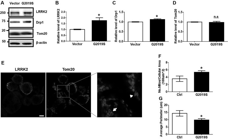
Figure 1.
Transfection of G2019S LRRK2 increases mitochondrial fission in BV2. (A-D) BV2 cells were transfected by empty vector (Vector) or myc-GS LRRK2 (GS) using Lipofectamine LTX for 6 h followed by 36 h incubation in the fresh growth medium. Cell lysates were subjected to Western blot analysis. Levels of Drp1 and Tom20 were normalized to α-tubulin levels. (E-G) GS LRRK2-expressing BV2 cells were analyzed by immunofluorescence using anti-LRRK2 [N241A/34] (Alexa-488, green) and anti-Tom20 (Texas Red, red). The morphology of representative mitochondria in GS LRRK2-expressing BV2 (GS) or non-expressing BV2 (Ctrl) is indicated by an arrowhead and an arrow, respectively. Statistical analyses were done using Student’s T-test with two-tailed p-value; *p < 0.05; n.s., not significant.

