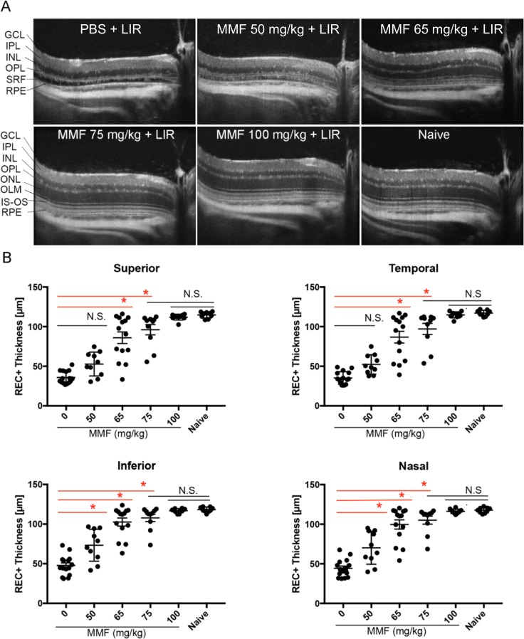
Figure 1.
MMF protects retinal structure from LIR. (A) Representative SD-OCT scans of the temporal retina. LIR caused ONL depletion and detachment between the RPE and OPL in PBS-treated mice, which was rescued by MMF as the dose increased from 50 to 100 mg/kg. OLM, outer limiting membrane; IS-OS, inner segment and outer segment junction; SRF, subretinal fluid. (B) Quantification of REC+ thickness in all four quadrants of retina showed that 50 and 65 mg/kg MMF were partially protective, whereas 75 and 100 mg/kg MMF provided full protection. Each dot represents the average REC+ thickness from the right and left eye of one mouse (n ≥ 9 in each group). Group average is shown as mean ± SE. N.S., nonsignificant (P > 0.05), *P < 0.05.

