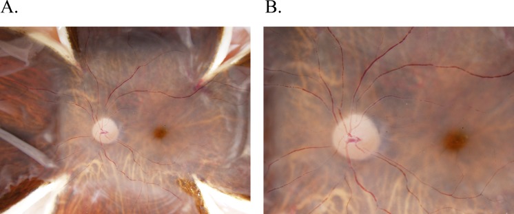
Figure 2.
Color fundus imaging in a normal donor eye. Following the butterfly dissection but prior to macular tissue isolation, retinal images are obtained for each eye at 0.7× (A) and 1.25× (B) magnifications using an Olympus SZX16 microscope camera illuminated with a Schott KL 1600 LED Fiber Optic Light Source Illuminator. Images are taken in the same orientation as done in a clinical setting for highest translational quality. As shown, detailed optic nerve, macula, fovea, and posterior pole vasculature can be seen.

