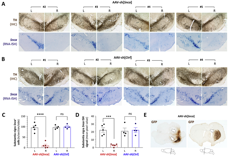
Figure 2: Loss of Snca mRNA expression in the substantia nigra and persistent transgene expression 52 weeks after AAV-sh[Snca] inoculation.

A: Photomicrographs of the substantia nigra are shown for animals #2 – #5 in the group receiving AAV-sh[Snca]. Each pair of images shows the substantia nigra on each side (labeled L and R) of the same section. Top row: immunohistochemistry (IHC) for tyrosine hydroxylase (TH; brown) showing the position and integrity of the substantia nigra. Bottom row: RNA in situ hybridization (ISH) for the Snca mRNA transcript (purple). IHC and ISH were performed on adjacent sections.
B: Similar to panel A, showing sections from each animal in the group receiving AAV-sh[Ctrl], Top row: IHC for TH (brown). Bottom row: RNA ISH for the Snca mRNA transcript (purple).
C: The number of Snca mRNA-expressing cells was counted on each side of the substantia nigra for three sections per animal. Individual data points show the mean value for each animal (AAV-sh[Snca] n=5; AAV-sh[Ctrl] n=4) and side; bars show the group mean ± 95% CI.
D: The Snca mRNA ISH signal was quantified by densitometry in the region corresponding to the substantia nigra on each side of three midbrain sections per animal. Individual data points show the mean value for each animal and side; bars show the group mean ± 95% CI.
E: Expression of the vector-encoded GFP transgene was determined in the midbrain (left) and forebrain (right) by IHC for GFP (brown), demonstrating persistent presence and expression of the AAV vector 52 weeks post-transduction. The cartoons below the micrographs indicate the planes of the sections shown.
Panels C and D were analyzed by 1-way ANOVA with Tukey’s post hoc test. ***p<0.0001, ****p<0.00001.
