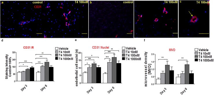
Fig 3. T4 stimulates angiogenesis in wounded human skin.
(a-c) To analyze angiogenesis, the number of CD31+ cells (red) and of CD31+ blood vessel cross-sections (lumina) (yellow arrow, c) per visual field were counted by immunofluorescence microscopy (at least 12 visual fields per skin fragment were evaluated). In addition, the intensity of CD31 IR was measured. Scale bars in a, b = 50μm, c = 200μm. (d) CD31 IR was significantly up-regulated by T4 at days 3 and 6. Immunoreactivity data was normalized to the control data as were (e, f) the number of CD31 +ve endothelial cell nuclei (CD31+/DAPI+ cells) and lumina per microscopic field. Number of independent experiments: n = 3 subjects (i.e. 1–2 punches per patient, per treatment group and per time point and at least 8 photomicrographs were analyzed per condition); data were pooled since the results trends in all three independent experiments were comparable). MVD: Microvessel density; ibFGF ab: inhibitory bFGF antibody.

