Fig. 1.
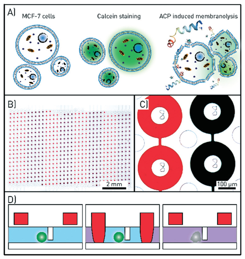
Assay and microfluidic chip design for single-cell drug testing. A) Schematics of the assay. Anticancer peptides (ACPs) are administered to MCF-7 cells loaded with calcein, which leaks out of the cells upon ACP-induced membranolysis. B) A microscopy image of the microfluidic channel with 612 chambers with central cell traps. The round valves that define the chambers when in the closed state are filled with red and black coloured solutions. Eight valve control lines are used to open and close the red and black subsets of the valves. C) Microscopy image of four microchambers and cell traps. The columns in between the chambers prevent the large channel and the chambers from collapsing. D) Schematic side view of one chamber. The actuation of the ring-valve (red) is used to isolate cell, ensure flow-free conditions during imaging, and precisely time the drug exposure.

