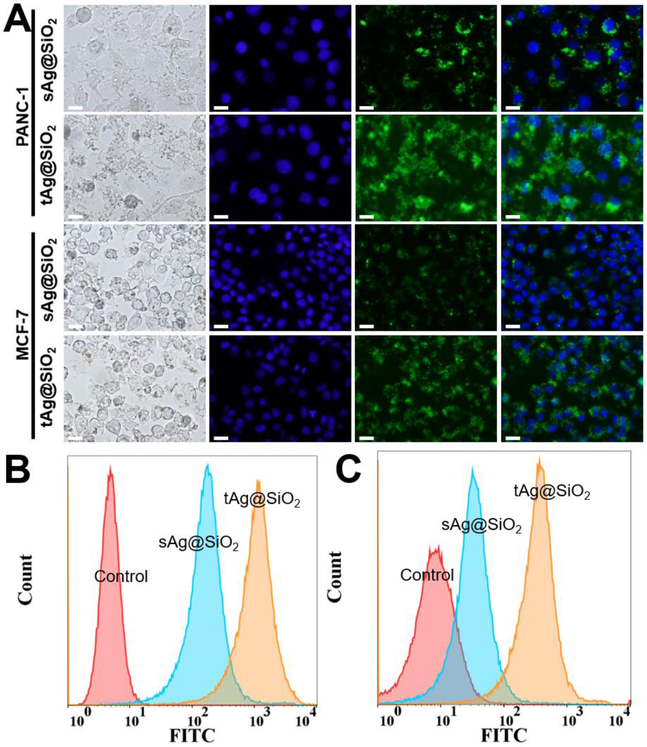
Figure 7.
Comparison of cellular internalization efficiency between the triangular and the spherical silver nanomaterials. (A) Bright-field and fluorescence images of PANC-1 and MCF-7 cells treated by sAg@SiO2 and tAg@SiO2 with a particle concentration of 100 μg/mL for 3 hours. The blue and green colors came from Hoechst 33342 and FITC-labeled nanoparticles. All scale bars denote a length of 20 μm. (B) and (C) are the flow cytometry analysis of core-shell nanoparticles-treated PANC-1 and MCF-7 cells, respectively.

