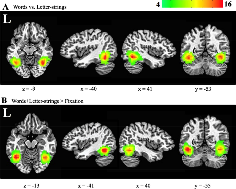
Figure 2.
VWFA and rVWFA subject coverage maps. Axial, left and right sagittal, and coronal views for (A) multivariate-defined (contrast: words versus letter-strings) and (B) univariate-defined (contrast: words+letter-strings > baseline) ROIs. All 22 participants are included in this map. Red represents voxels shared amongst more participants. Green represents voxels shared between fewer participants. The Talairach coordinates for the center of mass of these ROIs were: multivariate-defined VWFA (−40x, −53y, −10z), multivariate-defined rVWFA (41x, −52y, −10z), univariate-defined VWFA (−37x, − 55y, 10z), and univariate-defined rVWFA (39x, −55y, −11z).

