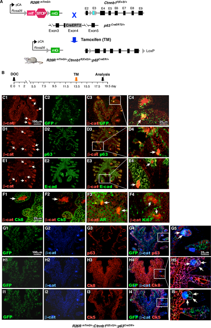
Figure 1. Conditional expression of stabilized β-catenin in p63-expressing cells at embryonic stage induces atypical cluster formation.

A. Schematic illustration of transgenic alleles and tamoxifen-inducible recombination events present in RosamTmG/+, Ctnnb1fl(Ex3)/+, and p63CreERT2/+ mice. The expression of either membrane-targeted tdTomato (mT) or membrane-targeted EGFP (mG) is regulated by the pCA promoter, consisting of a chicken β-actin core promoter with a CMV enhancer, through Cre/LoxP mediated recombination. B. Schematic representation depicting experimental timeline for labeling and analyzing UGS tissues during embryonic stage. Pregnant females received a single dose of tamoxifen on E13.5 and UGS tissues were isolated at E18.5. C-E. Immunofluorescent staining of UGS tissues from RosamTmG/+ Ctnnb1fl(Ex3)/+ p63CreERT2/+ embryos using antibodies against β-catenin (red), (C) GFP, (D) p63, (E) E-cad (green). F. Immunofluorescent staining of UGS tissues from RosamTmG/+ Ctnnb1fl(Ex3)/+ p63CreERT2/+ embryos using antibodies against β-catenin (red) and (F1) Ck8, (F2) Ck5, (F3) AR or (F4) Ki67 (green) G-I. Triple immunofluorescent staining of GFP (green), beta-catenin (blue) with (G) p63, (H) Ck8 or (I) Ck5 (red). Arrows (C-I) denote proliferative intraepithelial cell clusters expressing stabilized β-catenin (n=3).
