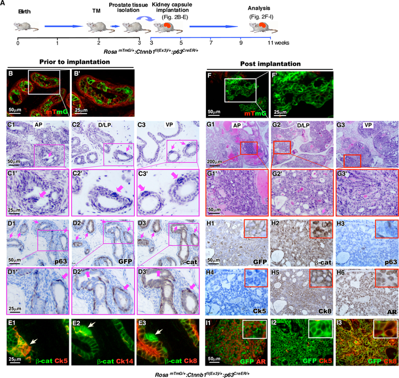
Figure 2. Conditional expression of stabilized β-catenin in prostatic prepubescent p63 expressing cells induces cell proliferation and oncogenic transformation.

A. Schematic representation of experimental design. RosamTmG/+ Ctnnb1fl(Ex3)/+ p63CreERT2/+ mice (n=6) received a single tamoxifen injection on P14. Prostate tissues were isolated and dissected into individual lobes one week later (P21). Half of each lobe was implanted under the kidney capsule of SCID mice, while the other half was preserved for histological and immunofluorescent analyses. Kidney grafts. B and F. mTmG fluorescence of (B-B’) P21 prostate tissues prior to implantation under the kidney capsule and (F-F’) graft tissues eight weeks after implantation in the kidney. C. H&E stained sections of different prostatic lobes, (C1-C1’) anterior prostate (AP), (C2-C2’) dorsolateral prostate (D/LP), and (C3-C3’) ventral prostate (VP) from RosamTmG/+ Ctnnb1fl(Ex3)/+ p63CreERT2/+ mice at P21. Immunohistochemical staining of sequential sections of the same tissues with (D1-D1’) p63, (D2-D2’) GFP or (D3-D3’) β-catenin antibodies (brown). Sections are counterstained with hematoxylin (blue). E. Immunofluorescent staining of stabilized β-catenin (green) with (E1) Ck5, (E2) Ck14 or (E3) Ck8 (red). G. H&E stained sections of graft tissues derived from different prostatic lobes. H. IHC staining of sequential sections of graft tissues with (H1) GFP, (H2) β-catenin, (H3) p63, (H4) Ck5, (H5) Ck8, or (H6) AR antibodies (brown). Sections are counterstained with hematoxylin (blue). I. Immunofluorescent images of graft tissues generated from RosamTmG/+ Ctnnb1fl(Ex3)/+ p63CreERT2/+ prostates stained with antibodies against GFP (green) and (I1) AR, (I2) Ck5, or (I3) Ck8 (red).
