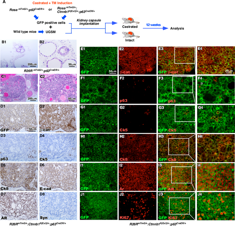
Figure 5. Stabilization of β-catenin in sensitized p63-expressing cells resulted in oncogenesis in tissue recombinants.

A. Schematic depicting preparation of recombinants and timeline for kidney capsule transplantation. RosamTmG/+ p63CreERT2/+ or RosamTmG/+ Ctnnb1fl(Ex3)/+ p63CreERT2/+ mice were castrated at the age of eight weeks, injected with TM at the age of twelve weeks, and sacrificed eight weeks later. Prostate tissues were isolated and cells were sorted for GFP expression using FACS. GFP-positive cells were implanted under the kidney capsule of intact SCID mice along with wild-type urogenital sinus mesenchyme tissue. Tissue grafts were isolated twelve weeks later (n=3). B-C. H&E stained sections of tissue graft recombinants made of UGSM and GFP-positive epithelial cells from the prostates of either (B) RosamTmG/+ p63CreERT2/+ or (C) RosamTmG/+ Ctnnb1fl(Ex3)/+ p63CreERT2/+ mice. D. IHC staining of sequential sections of tissues grafts from RosamTmG/+ Ctnnb1fl(Ex3)/+ p63CreERT2/+ mice using antibodies against (D1) GFP, (D2) β-catenin, (D3) p63, (D4) Ck5, (D5) Ck8, (D6) E-cad, (D7) AR or (D8) Synaptophysin (brown). Sections are counterstained with hematoxylin (blue). E-J. Immunofluorescent staining of tissue recombinants generated from RosamTmG/+ Ctnnb1fl(Ex3)/+ p63CreERT2/+ mice using antibodies against GFP (green) and (E) β-catenin, (F) p63, (G) Ck5, (H) Ck8, (I) AR, or (J) Ki67 (red).
