Figure 2.

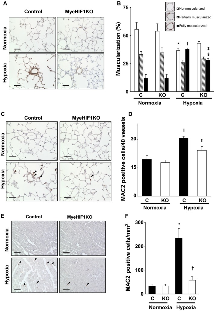
Muscularization in the distal pulmonary arteries after 3 weeks under hypoxic conditions. Macrophage infiltration into the lung after 3 days. (A) Representative α‐SMA‐stained mouse lung tissue (brown) of control mice or MyeHIF1KO mice in normoxia or hypoxia (10%, 3 weeks). Scale bars are 50 μm. (B) The percentage of hypoxia‐induced muscularization of distal pulmonary arteries (<70 μm diameter) by characterization as nonmuscularized, partially muscularized, and fully muscularized. Forty vessels were counted per sample. (C) Representative microphotographs of lung immunohistochemistry for MAC2‐positive macrophages (brown) after 3 days exposure to hypoxia (10%) or normoxia are shown. Scale bars are 50 μm. The arrowheads indicate representative MAC2‐positive cells. (D) The number of MAC2‐positive macrophages around the pulmonary arteries of 40 vessels from each sample. (B), n = 6 (C+normoxia), 6 (KO+normoxia), 7 (C+hypoxia), 6 (KO+hypoxia). (D), n = 8 (C+normoxia), 7 (KO+normoxia), 8 (C+hypoxia), 7 (KO+hypoxia). * P < 0.05 versus nonmuscularized for control mice in normoxia. † P < 0.05 versus fully muscularized for control mice in normoxia. ‡ P < 0.05 versus fully muscularized for KO mice in normoxia. § P < 0.05 versus fully muscularized for control mice in hypoxia. MyeHIF1KO mice. || P < 0.05 versus control mice in normoxia. ¶ P < 0.05 versus control mice in hypoxia. C, control mice; KO, MyeHIF1KO mice. (E) Representative microphotographs of immunohistochemical analysis for MAC2‐positive macrophages in the right ventricle (brown) after 3 weeks’ exposure to hypoxia (10%) or normoxia are shown. Scale bars are 50 μm. The arrowheads indicate MAC2‐positive cells. (F) The number of MAC2‐positive cells per square millimeter from each sample are counted and indicated in bar graphs. n = 6 (C+normoxia), 7 (KO+normoxia), 6 (C+hypoxia), 7 (KO+hypoxia). * P < 0.05 versus control mice in normoxia. † P < 0.05 versus control mice in hypoxia. C, control mice; KO, MyeHIF1KO mice.
