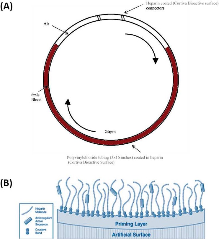
Fig. 1.
Chandler loop designed to mimic portal vein blood flow. a Polyvinyl chloride tubing (3 × 16 in.) was filled with 6 ml ABO-matched blood and hepatocytes. Loops were closed into circuits with heparin-coated polystyrene connectors. Tubing loops were rotated at 24 rpm and incubated at 37 °C for 0, 15, 30 and 60 min. b Schematic of Cortiva® BioActive Surface End Point Attached Heparin (http://www.medtronic.com/us-en/healthcare-professionals/products/cardiovascular/cardiopulmonary/cortiva-bioactive-surface.html). This method of anti-coagulation attaches heparin molecules via covalent bonds to amine groups on the prepared material surface. The aldehyde group on the heparin molecule is bound to the surface and the remainder of the molecule, including the active binding sequence is free to interact with the blood such as antithrombin

