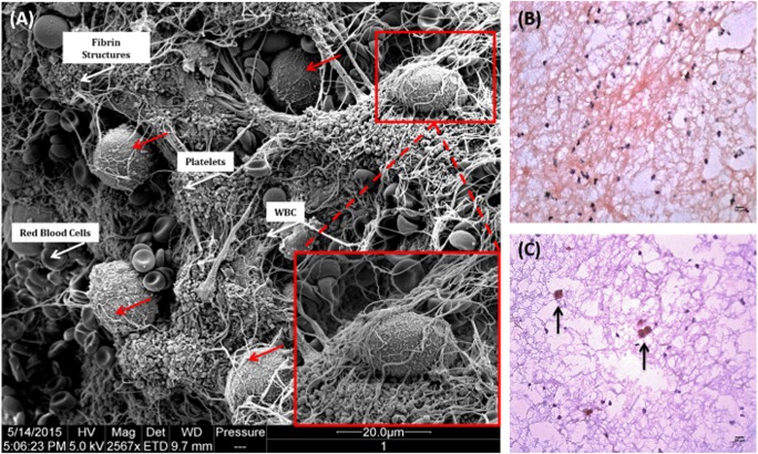
Fig. 3.
Primary human hepatocytes trigger large thrombus formation when in contact with ABO-matched blood. a Thrombi that formed within the Chandler loop model were fixed in 2.5% glutaraldehyde and prepared for electron microscopy. Hepatocytes became entrapped within thrombi, shown by red arrows and magnified inset image. Red blood cells, white blood cells, platelets and fibrin deposits are all visible within the thrombus (white arrows). Samples were examined and recorded using a FEI Quanta 200F field emission scanning electron microscope operated at 35 kV in high vacuum mode. Eight micrometre sections were cut and stained with a monoclonal mouse anti-human hepatocyte antibody (Clone OCH1E5), followed by a dual HRP polymer secondary antibody and counterstained with haematoxylin and eosin. b Thrombus from control loop containing blood only. c Thrombus from loop containing ABO-matched blood and hepatocytes. × 100 total magnification

