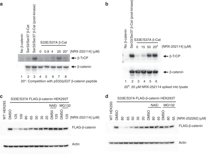
Fig. 6.
Enhancers degrade S33E/S37A phosphomimetic β-catenin protein in cells. a Purified Myc/FLAG-tagged β-catenin protein (as indicated) was mixed with HEK293T cell lysates in the presence of varying amounts of NRX-252114 and immunoprecipitated using anti-myc beads. The samples were resolved on SDS-PAGE and analyzed by immuno-blotting with either C-terminal β-catenin or β-TrCP antibody. In lane 3, 2 μM β-catenin was phosphorylated with the mix of 1 μM GSK3, 200 nM CK1 and 50 nM Axin prior to its addition to the lysate. In lane 8, 6.4 μM pSer33/pSer37 β-catenin peptide was spiked into the lysate prior to the addition of β-catenin protein. b HEK293T cells without (lanes 1 and 2) or with overexpression of S33E/S37A Myc/FLAG-tagged β-catenin (lanes 3-6) were treated with NRX-252114 for 6 h (as indicated). Cells were then lysed, β-catenin was immunoprecipitated using anti-myc beads and probed for either β-catenin or β-TrCP (as described above). In lane 2, cell lysate was spiked with phosphorylated WT Myc/FLAG-tagged β-catenin protein, whereas in lane 6, the lysate was spiked with 20 μM NRX-252114 as positive controls. c HEK293T cells stably expressing Myc/FLAG-tagged S33E/S37A β-catenin were treated with varying concentrations of NRX-252114 (as indicated) for 6hrs and examined for S33E/S37A β-catenin levels by western analysis using anti-FLAG antibody. For control, Actin levels were monitored using anti-Actin antibody. For co-treatments, either 5 μM of Nedd8 E1 inhibitor (NAEi) or 10 μM of proteasome inhibitor (MG132) were added to cells 30 min prior to the compound treatment. d Same as Fig. 6c, except NRX-252262 was used

