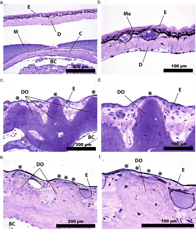
Figure 3.
Dermal ossification in pumpkin toadlets. Photomicrographs of transverse histological sections of heads of Brachycephalus hermogenesi (a,b), B. ephippium (c,d) and B. pitanga (e,f). BC: brain cavity, C: cranial bone, D: dermis, DO: dermal ossification, E: epidermis, M: muscle, Me: melanophore. Asterisks indicate points where the fluorescent ossified tissue is visible through the thin epidermis in live specimens. B. hermogenesi lacks dermal ossification and a layer of melanophores is present, in contrast to B. ephippium and B. pitanga.

