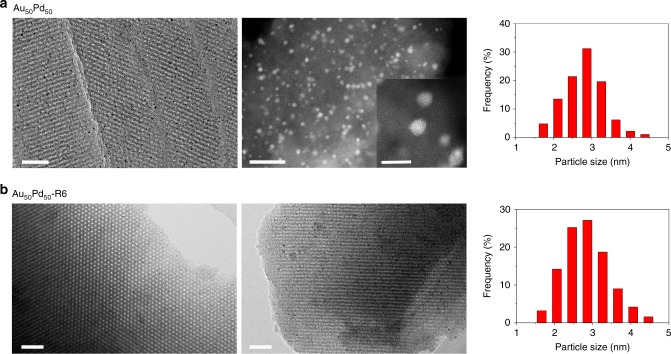
Fig. 2.
TEM micrographs. a Representative TEM image of the ultrathin sections (Left, the scale bar of 50 nm) and AC-STEM image (Middle, the scale bar of 20 nm) for the fresh Au50Pd50. Inset Middle figure is the AC-STEM image with a high magnification, and the scale bar is 5 nm. b TEM images of the recycled Au50Pd50-R6 after six catalytic runs viewed along the [001] and [110] directions. The scale bar is 50 nm. Particle size distributions (Right (a) and (b)) were determined with at least 200 nanoparticles

