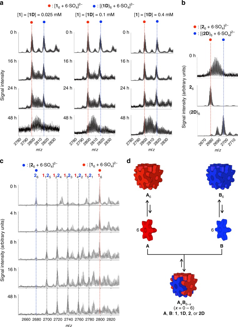
Fig. 2.
Monitoring of scrambling of GSAs between the nanocubes by ESI-TOF mass spectrometry and scrambling mechanism. a The scrambling of GSAs between 16 and (1D)6 was monitored in different concentrations. Right after the mixing of aqueous solutions of 16 and (1D)6, strong signals for the homoleptic nanocubes (indicated by solid blue and red circles) were observed. These signals became weak with time and new signals assigned to heteroleptic nanocubes appeared in between the two signals. The scrambling takes place faster in lower concentration of the nanocubes. b The scrambling of GSAs between 26 and (2D)6. The scrambling was completed right after the mixing of 26 and (2D)6, which is much faster than the scrambling between 16 and (1D)6. c The scrambling of GSAs between 16 and 26 was monitored after mixing of aqueous solutions of 16 and 26. The conversion of the less stable nanocube (26) into heteroleptic nanocubes, 1x26−x (x = 1–5), is faster than that of 16. d The scrambling of GSAs between the nanocubes A6 and B6 takes place through the exchange of monomer GSAs dissociated from the nanocubes. After the formation of heteroleptic nanocubes AxB6−x (x = 1–5), the scrambling of AxB6−x (x = 1–5) with the less stable homoleptic nanocube takes place faster than with the more stable one

