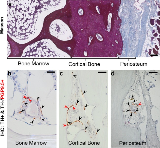
Figure 1.
Illustration of the method used for the analysis of nerve profiles in human bone. (a) Masson’s trichrome stained section showing the overall compartmentalization of the iliac crest biopsy into bone marrow, cortical bone, and periosteum. Sections were sequentially immunostained for tyrosine hydroxylase (TH, black arrows) and protein gene product 9.5 (PGP9.5, red arrows). The images illustrate this immunostaining within the bone marrow (b), cortical pore (c) and periosteum (d). The sequential immunostaining allows discrimination between the TH+ nerve profiles with sympathetic nerve fibers (brown) and the TH−/PGP9.5+ nerve profiles without sympathetic nerve fibers (red). Scale bars are 200 µm (a) and 50 µm (b–d).

