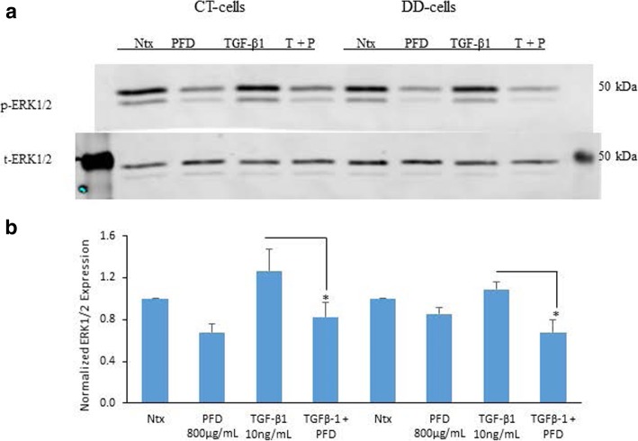
Fig. 2.
Pirfenidone significantly reduced TGF-β1-stimulated phosphorylation of ERK1/2 in both CT- and DD-derived fibroblasts. a CT- and DD-cord derived fibroblasts derived from three different patient samples (N = 3/group) were maintained in α-MEM medium containing 0.1% dialyzed FBS for 24 h. After 24 h, cells were either left as controls or treated with PFD (800 μg/ml) in the presence or absence of TGF-β1 (10 ng/ml) for 15 min. Cell lysates were subjected to Western blot analyses to determine the expression of phosphorylated ERK1/2. b Densitometry results are reported as the ratio of phosphorylated ERK1/2 protein level to GAPDH expression. Values are means ± standard error mean (SEM) of three independent studies from each of CT- and DD- derived fibroblasts. Shown here is a representative image of Western blots from three different cultures of CT- and DD-cord derived fibroblasts, each showing similar results. *p < 0.04, . Ntx; No treatment, PFD- Pirfenidone, TGF-β1, T + P; TGF- β1 + Pirfenidone

