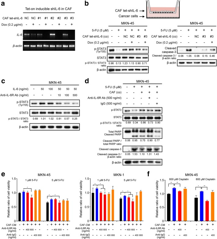
Fig. 3.
Suppressive effect of interleukin-6 (IL-6) inhibition on the cancer-associated fibroblast (CAF)-induced resistance to 5-fluorouracil (5-FU). a Reverse transcription (RT)-PCR analysis showing the expression of IL6 and ACTB mRNA in CAFs transfected with three different tet-on inducible IL6 shRNAs vectors or a negative control vector [38]. Dox indicates doxycycline. b Schematic figure depicting the transwell co-culture system for tet-on IL6 shRNA-transfected CAFs and gastric cancer cells. Western blot analysis shows the expression of the apoptotic markers cleaved PARP, caspase-3, and phosphorylated STAT3 in the lysate of MKN-45 cell cultures in the lower chamber after doxycycline (0.2 μg/ml) treatment of CAFs transfected with the tet-on IL6 shRNA or negative control (NC) vector in the upper chamber. c Western blot analysis showing the expression of the indicated proteins in cells treated with human recombinant IL-6 combined with and without tocilizumab treatment. d Western blot analysis showing the expression of the indicated proteins in the lysates from MKN-45 and MKN-1 cells after 5-FU (5 μM) treatment with and without CAFs and subsequent treatment with tocilizumab (500 ng/ml) or negative control IgG (500 ng/ml). e Ez-cytox tests showing the relative ratio of the viability of MKN-45 and MKN-1 cells treated with 1 μM or 5 μM of 5-FU after the addition of tocilizumab (400 and 800 ng/ml) or control IgG (400 and 800 ng/ml). f Ez-cytox tests demonstrating the relative ratio of cell viability in MKN-45 cultures treated with 600 μM or 800 μM cisplatin after the addition of tocilizumab (400 ng/ml) or control IgG (400 ng/ml). The graphs show the mean (± SEM) ratios of cell viability. *P < 0.05 and **P < 0.001, according to Mann-Whitney test

