Fig. 1.
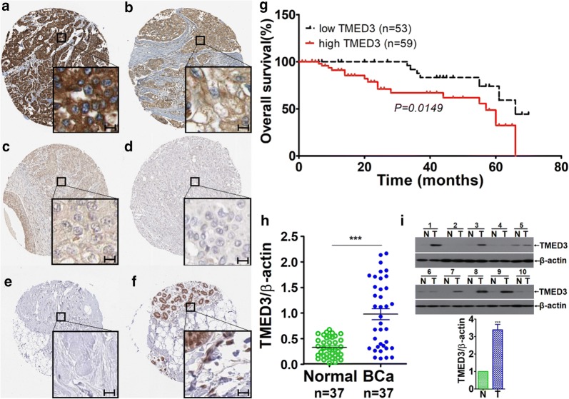
Up-regulated TMED3 significantly is associated with unfavorable overall prognosis in breast cancer. a Strong immunostaining of TMED3; b moderate staining of TMED3; c weak staining of TMED3; d negative staining of TMED3 in breast cancer tissue; e negative staining of TMED3 in normal breast tissue; f moderate staining of TMED3 in the duct of normal breast tissue. Scale bar stands for 5 µm; magnification fold for overview of dots was ×5, whereas ×40 for insets. Shown were the representative figures selected from candidates. g Kaplan–Meier survival curve of TMED3 expression in patients with breast cancer, divided into high and low group according to the status of TMED3 expression, with each group having 59 and 53 cases respectively. Log-rank test was used to analyze the difference of overall survival. h qRT-PCR detection of TMED3 expression on mRNA level in 37 cases of breast cancer and its matched normal control tissues. ***p < 0.001 compared with control normal group using independent sample T-test; i western-blot analysis of TMED3 in the fresh breast cancer tissues, totaling 10 paired cases. T tumor tissue, N normal breast tissue. The molecular weight (MW) of TMED3 was around 25 kDa, β-actin, as internal loading control whose MW was observed to be about 42 kDa. Quantitative assay was performed using Image J software (NIH, Bethesda, USA), ***p < 0.001 relative to control group using independent sample T-test

