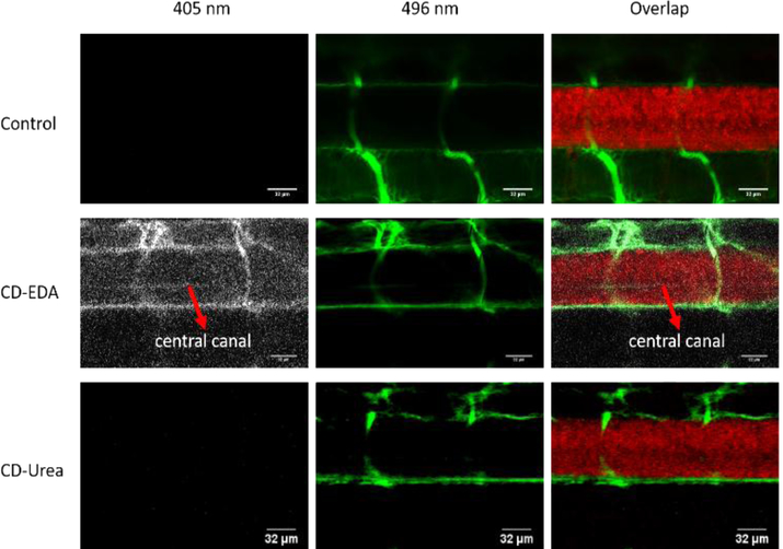
Figure 3:
Confocal microscopic images of a six-day-old, transgenic zebrafish larva expressing mcherry (585 nm) in the central nervous system. The larvae were injected with either 10,000 MW fluorescein dextran dye (496 nm) alone (control, top row), or a combination of dye and CD-EDA (second row) or a combination of dye and CD-Urea (third row). Fluorescence from CDs (405 nm) that cross the blood brain barrier can be seen in the central canal that is highlighted with the red arrows.

