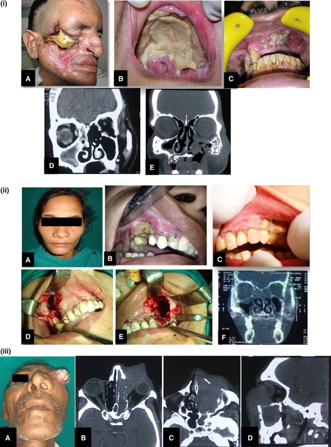
Fig. 4.
Clinico-radiologic presentations of FRS. (i) A Extensive cheek necrosis, B palatal necrosis, C exposed necrotic bone on intraoral examination, D CT scan of a patient diagnosed with Allergic FRS. Coronal section showing hyperdensity of mucin within right ethmoid and maxillary sinuses, E coronal CT image showing mixed lytic sclerotic changes with bony destruction involving left zygoma and walls of left maxillary sinus. Also there is soft tissue attenuation with air loculi. (ii) A A patient at clinical presentation with very mild diffuse bilateral swelling, B, C intraoral pictures showing exposed necrotic bone and generalized mobility of maxillary teeth stabilized with Essig’s wiring, D, E intraoperative pictures for sequestrectomy and curettage, F CT scan image shows mucosal thickening of the maxillary sinuses with erosion of bone. (iii) A A clinical presentation of case of CIFRS with extensive orbital involvement mimicking malignancy, B, C, D radiographic picture of bone erosion localized to the area of extra-sinus component of the disease more extensive than intra-sinus component

