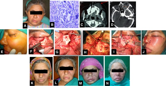
Fig. 5.
Photographic series of management of a patient with FRS. A preoperative clinical presentation of immunocompetent patient showing diffuse swelling on left side of face and orbital involvement. B Histopathology showing PAS-positive wall of a negatively stained section showing fungal hyphae(× 400), C Axial CT scan shows diffusely enhancing mass (arrow) involving the left maxillary sinus and the subcutaneous plane of the left cheek and upper lip, D Axial CT scan showing left eye proptosis caused by a mass involving the temporal fossa and orbit extending to involve the ethmoid air cells with bone erosion(arrows), E Weber- Fergusson incision, F Zygomatic swing osteotomy to get an access to orbital floor, G Zygoma swung laterally, H Surgical debridement extending to ethmoid sinus, I Realignment, J Closure, K 2 months post operative picture after a course of Amphotericin B, L 1 year postoperative patient had vision loss and was given Voriconazole after which disease no longer progressed M 5 years post treatment patient had another relapse, N Patient responded well to a course of Voriconazole again

