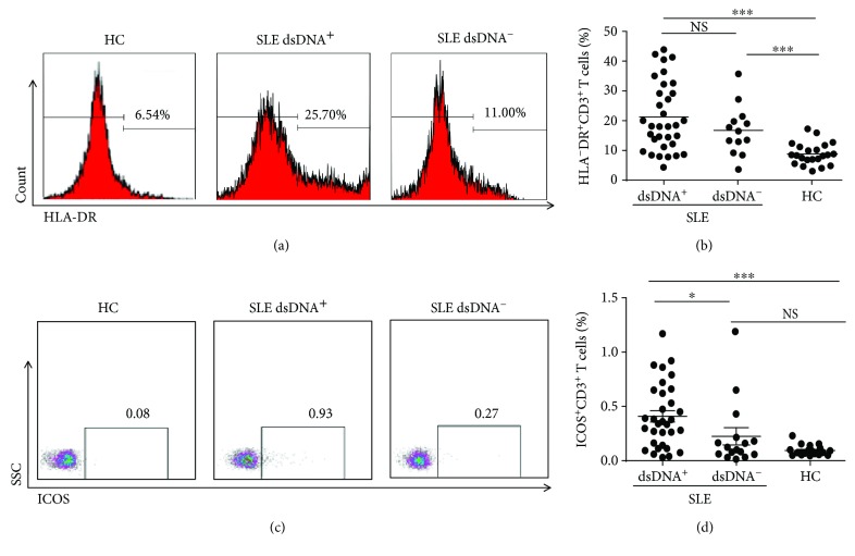
Figure 1.
Frequencies of HLA-DR+CD3+ and ICOS+CD3+T cells in peripheral blood. Peripheral blood mononuclear cells (PBMCs) from double-stranded DNA (dsDNA)+ systemic lupus erythematosus (SLE) patients (n = 23), dsDNA−SLE patients (n = 17), and healthy control subjects (n = 22) were harvested and stained with appropriate flow antibodies, and the expression of HLA-DR+CD3+ and ICOS+CD3+ cells was analyzed by flow cytometry. (a, b) Representative dot plots of HLA-DR+CD3+ T cells (a) and the summarized graph (b) are displayed. (c, d) Representative dot plots (c) and pooled data (d) of ICOS+CD3+ T cells are displayed. Each dot in (c, d) represents one subject. ∗ P < 0.05, ∗∗ P < 0.01, ∗∗∗ P < 0.001, NS: no statistical significance.

