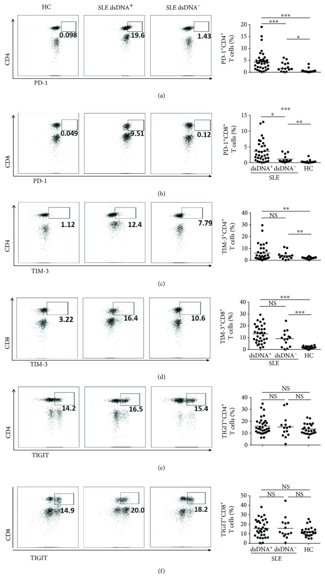
Figure 2.
Expression levels of PD1, TIM3, and TIGIT on blood T leukocytes. PBMCs from dsDNA+ systemic lupus erythematosus (SLE) patients (n = 23), dsDNA−SLE patients (n = 17), and control subjects (n = 22) were harvested and stained with appropriate flow antibodies, and the expressions of PD-1, TIM-3, and TIGIT on CD3+ T cells were detected by flow cytometry. The flow figures and pooled data for PD-1+ CD4+ T cells (a), PD-1+ CD8+ T cells (b), TIM-3+CD4+ T cells (c), TIM-3+ CD8+ T cells (d), TIGIT+ CD4+ T cells (e), and TIGIT+ CD8+ T cells (f) are shown. Each dot in the statistical graphs represents an individual subject. ∗ P < 0.05, ∗∗ P < 0.01, ∗∗∗ P < 0.001, NS: no statistical significance.

