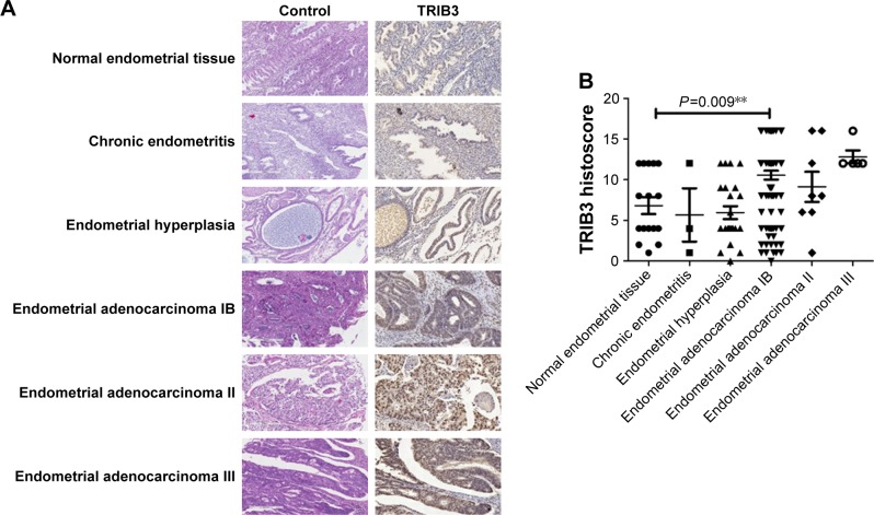
Figure 1.
Immunohistochemical analysis of the expression of TRIB3 in control tissue and tissues with endometritis, endometrial hyperplasia, and endometrioid adenocarcinoma (stages IB, II, and III).
Notes: (A) Representative examples of TRIB3 staining. TRIB3 staining was mainly localized in the cytoplasm. (B) Quantitation of immunohistochemical data. **P<0.01 by ANOVA.

