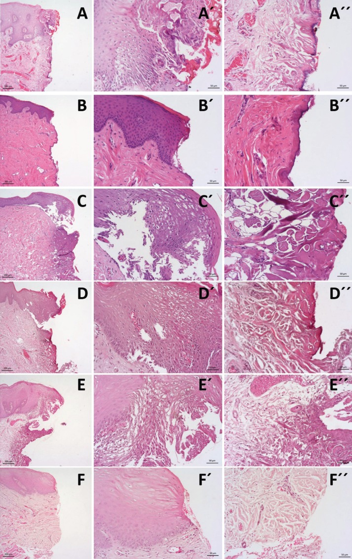
Figure 1.
Histological images (haematoxylin and eosin staining) at magnification of 5x and 20x for epithelium view (´) and connective view (´´) of the surgical margins of tissue samples submitted to excision by the 6 groups of instruments: A –CO2 Laser; B –Er:YAG Laser; C –Diode Laser; D – Nd:YAG laser; E – electrical surgical scalpel; F – cold scalpel.

