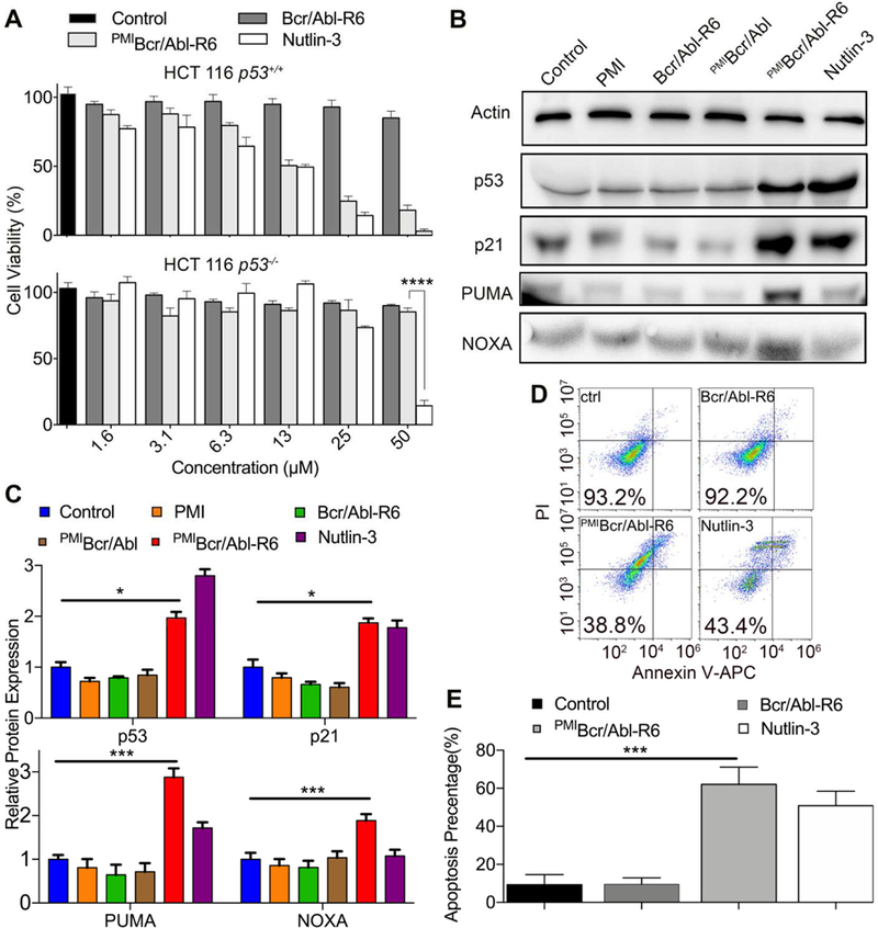
Fig. 5. PMIBcr/Abl-R6 kills HCT116 p53+/+ tumor cells in vitro by reactivating the p53 pathway.

(A) Cell viability of HCT116 p53+/+ and HCT116 p53−/− cells (3×10³ cells/well in McCoys’s 5A medium with 10% FBS) 48 h after treatment with varying concentrations of Bcr/Abl-R6, PMIBcr/Abl-R6 and Nutlin-3. Following a 2-h incubation with CCK-8 reagents, absorbance values at 450 nm were measured on a microplate reader, and percent cell viability was calculated as (Atreatment−Ablank)/(Acontrol−Ablank) × 100%. The data are the means of three independent assays. Except for HCT116 p53−/− cells treated by PMIBcr/Abl-R6 and Nutlin-3 at 50 µM (****, p<0.0001), no statistically significant difference in activity between PMIBcr/Abl-R6 and Nutlin-3 was found. (B) Representative Western blotting analysis of p53, p21, PUMA, NOXA in HCT116 p53+/+ cells (2×10⁴ cells/well) 48 h after treatment with PMI, PMIBcr/Abl, PMIBcr/Abl-R6, Bcr/Abl-R6 and Nutlin-3 at 12.5 µM each, normalized to β-actin. The primary antibodies were from Santa Cruz Biotechnology (p53), Calbiochem (p21, PUMA and NOXA) and Sigma-Aldrich (β-actin), and secondary antibodies conjugated with horseradish peroxidase from Calbiochem. (C) Quantitative Western blotting analysis (via Image J software) of HCT116 p53+/+ cells treated with PMI, Bcr/Abl-R6, PMIBcr/Abl, PMIBcr/Abl-R6 and Nutlin-3 at 12.5 µ M for 48 h. T-test was performed for statistical analysis, * standing for p<0.05, *** for p<0.001. The data are the means ± SD of three independent Western blotting assays. (D) Representative data on apoptosis of HCT116 p53+/+ cells 48 h after treatment with Bcr/Abl-R6, PMIBcr/Abl-R6 and Nutlin-3 as analyzed by flow cytometry. Cells were seeded in a 12-well plate with a density of 20,000/well and treated with 12.5 µM PMIBcr/Abl-R6, Bcr/Abl-R6 or 10 µ M Nutlin-3. Apoptosis was detected using a standard apoptotic kit from Biolegend, including APC labeled anti-annexin V antibody and a propidium iodide solution. (E) Statistical analysis of apoptosis of HCT116 p53+/+ cells quantified by flow cytometry. Three independent FACS assays were performed, and data are shown as the means ± SD (n=3). p values were calculated by t-test (***, p<0.001).
