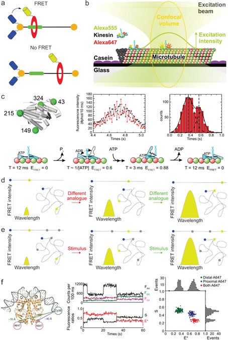
Fig. 4.
Determining molecular conformations using FRET. (a) Schematic depiction of a rotaxane consisting of a rotor (red) and stator (black and green). By labeling the rotor and stator with a FRET pair, the rotor’s position along the stator can be monitored. Image based on ref62. (b) Using FRET to monitor kinesin movement along a microtubule by labeling the two domains of kinesin (blue) with a fluorophore (green and red). Image adapted from ref65. (c) Schematic of a kinesin motor domain functionalized with 4 cystine residues facilitating site-specific labeling with fluorophores. By monitoring the fluorescence intensity of a FRET-pair labeled kinesin over time (black line donor fluorescence intensity, red line acceptor fluorescence intensity), two populations of FRET efficiency could be observed corresponding to different stages in kinesin’s step. By repeating such experiments using differently labeled kinesin analogues, detailed information of kinesin’s gait could be obtained. Images b and c are both adapted from ref65. Copyright 2009, National Academy of Sciences. (d) In a typical FRET conformational mapping experiment, various analogues with fluorophores at predefined positions are synthesized and analyzed. The obtained FRET intensities allow subsequent reconstruction of the molecular conformation. Blue and yellow dots represent the donor and acceptor fluorophore, respectively; black lines represent the molecule in its stretched and folded conformation. (e) In switchable FRET the molecule of interest is labeled with one donor fluorophore (blue) and multiple stimuli responsive acceptor fluorophores (yellow when on, gray when off). By sequentially measuring the FRET efficiency between the acceptor and each donor fluorophore, multiple distances can be determined using the same compound. (f) Example of switchable FRET results, used to determine molecular conformation. The following fluorescence intensities were monitored: FAA = acceptor emission upon acceptor irradiation, FDD = donor emission upon donor irradiation, FDA = acceptor emission upon donor irradiation. The acceptor fluorophores were switched in the following manner: 5 – 20 seconds, only proximal A647 on; 20 – 35 seconds, both acceptors on; 35 – 60 seconds, only distal A647 on). Image adapted with permission from ref68. Copyright 2010, Nature Publishing Group.

