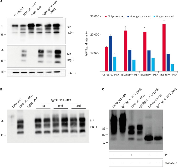
Fig. 2. Abnormal PrP detection in ME7-infected C57BL/6J and TgSShpPrP mice. (A) Western blot analysis for PrPc and PrPSc in ME7-infected mice at 2nd passage. 20 μg of total protein were loaded into each well. For PrPSc detection, the brain homogenates were digested with 7 µg/mL of PK. PrPc and PrPSc were detected using mAb 7A12. Relative densities of un-, mono-, and di-glycosylated forms of PrPSc obtained from ME7-infected mice obtained using image J analyzer. (B) Western blot analysis for PrPc and PrPSc in ME7-infected mice at 1st, 2nd, and 3rd passages. Total protein (20 μg) was loaded into each well. In order to detect PrPSc, the brain homogenates were digested with 7 µg/mL of PK. The PrPc and PrPSc were detected using mAb 7A12. (C) To confirm the electrophoretic mobility of PrPSc from ME7-infected C57BL/6J and TgSShpPrP mice, brain homogenates were treated with PK and PNGase F prior to western blotting with mAb 7A12.
PrP, prion protein; TgSShpPrP, transgenic mouse line carrying the Suffolk sheep PrP gene; PrPc, cellular PrP; PrPSc, scrapie PrP; PK, proteinase K; PNGase F, peptide N-glycosidase F.

