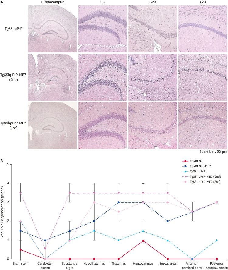
Fig. 3. H&E staining and severity of neurodegeneration associated vacuolation in TgSShpPrP-ME7 infected brains. (A) H&E staining of age-matched control TgSShpPrP and terminally ill ME7-infected mice showing the dentate gyrus, CA3, and CA1 regions of the hippocampus. The hippocampus was viewed at a low magnification (4×); all other regions were viewed at a higher magnification (40×). (B) Mean vacuolation scores and standard errors of the means from brains of ME7-infected C57BL/6J and TgSShpPrP mice and age-matched control mice.
PrP, prion protein; H&E, hematoxylin and eosin; TgSShpPrP, transgenic mouse line carrying the Suffolk sheep PrP gene; DG, dentate gyrus; CA, cornu ammonis.

