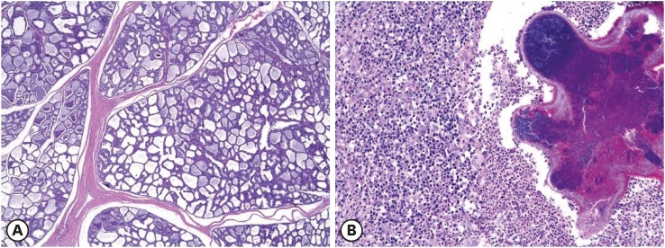
Fig. 1. Histopathological analysis of mammary lesions in sows. (A) Mammary gland hyperplasia. The number of mammary gland ducts, as well as the number of their epithelial cells, are elevated (H&E stain, 40×). (B) Mastitis. Inflammatory cells composed of lymphocytes, macrophages, and neutrophils congregate around a central bacterial focus (H&E stain, 200×).
H&E, hematoxylin and eosin.

