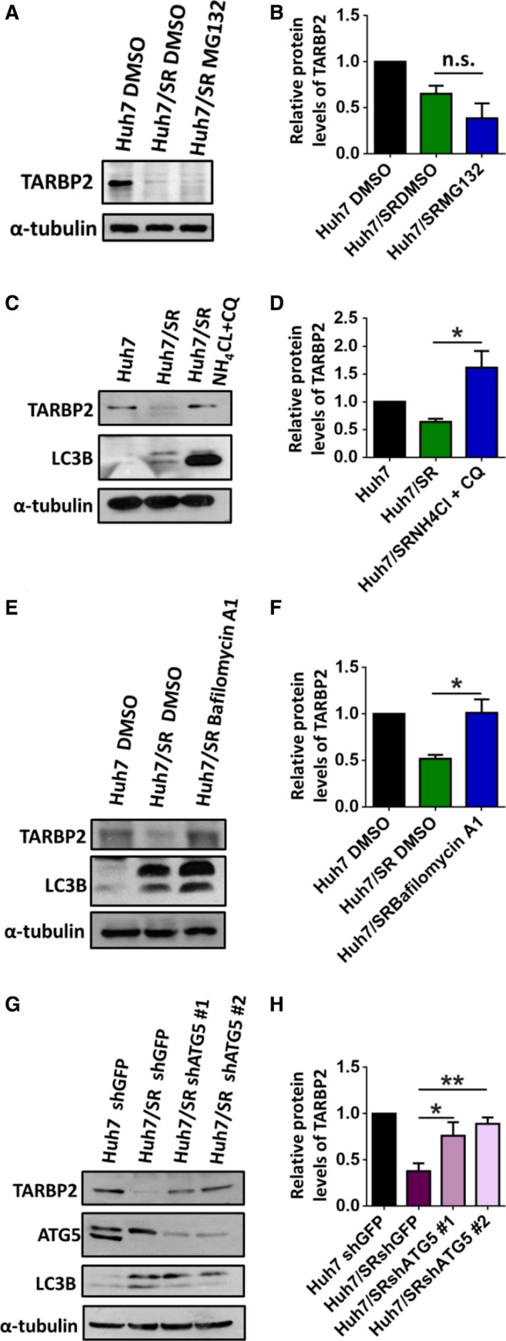
Figure 4.

The TARBP2 protein is destabilized through the autophagic–lysosomal pathway. Effects of the proteolytic pathways on TARBP2 downregulation in HCC/SR cells. (A, B) Huh7/SR cells were treated with MG132 (5 μm) for 24 h. (C, D) Huh7/SR cells were treated with NH 4Cl (10 mm) and CQ (200 μm) for 48 h. (E, F) Huh7/SR cells were treated with BFA (100 nm) for 48 h. (G, H) ATG5 was knocked down in the Huh7/SR cells. The TARBP2, ATG5, and LC3B proteins were quantified via western blot analysis. Data are presented as the mean ± SEM from at least three independent experiments. Multigroup comparisons were analyzed by one‐way ANOVA with Tukey's post hoc test. P values < 0.05 were considered statistically significant. *P < 0.05; **P < 0.01.
