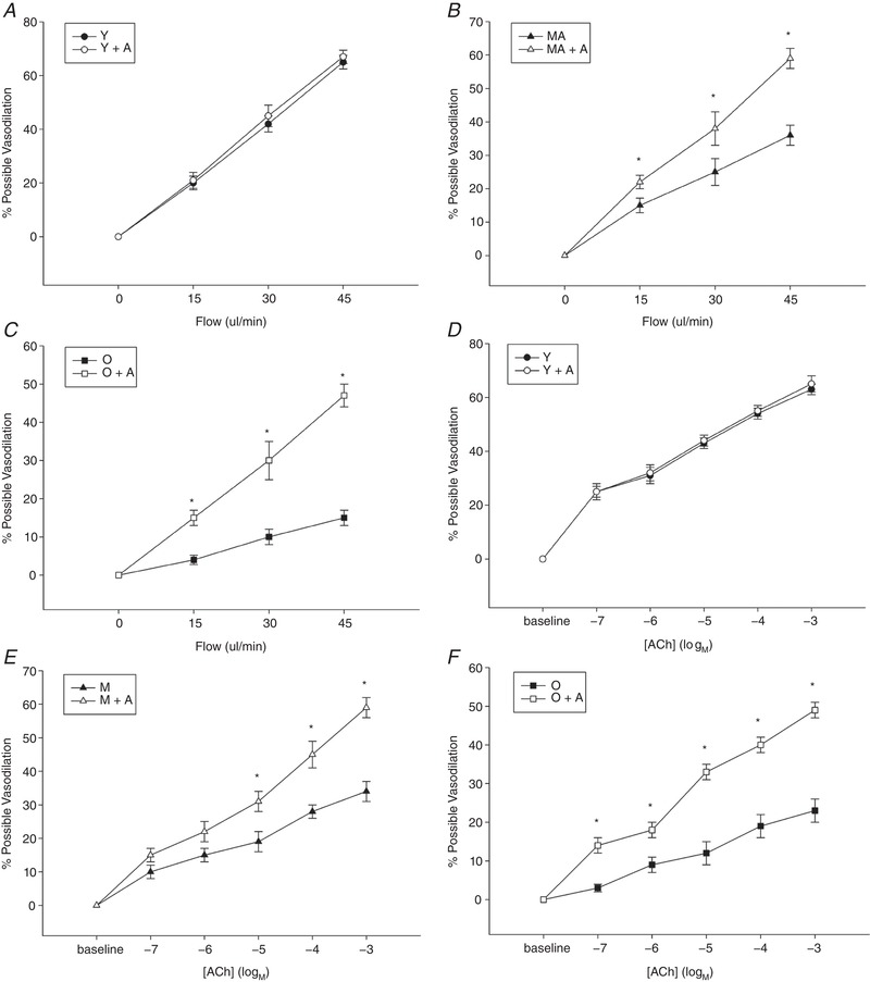
Figure 3. Skeletal muscle feed arteries with and without adropin incubation evoked by flow and ACh.

The vasodilator dose–response curves of skeletal muscle feed arteries from Young, Middle Aged and Old subjects with and without adropin incubation evoked by flow (A–C) and ACh (D–F). Data are expressed as the mean ± SE. n = 10 Young, 10 Middle Aged and 16 Old subjects. *Significant difference between with and without adropin incubation, P < 0.05.
