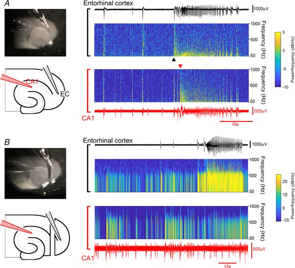
Figure 2. Tonic–clonic activation of CA1 can spread from entorhinal cortex.

A, ventral horizontal brain slice, in which tonic–clonic activity spreads from entorhinal cortex into CA1. The spectrograms show that the sustained (tonic) high frequency component occurs first in the entorhinal cortex. B, recording from the same slice, following sectioning of the temporoammonic pathway. Note the tonic–clonic‐like event in entorhinal cortex, but not in CA1. [Color figure can be viewed at wileyonlinelibrary.com]
