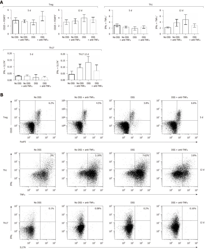
Figure 4.
T cells subsets characterization in presence of an anti-tumor necrosis factor α agent. T cells were isolated from mesenteric lymphnode of mice and CD4+ cells were studied by flow cytometry. In particular, CD4+ regulatory T lymphocytes characterization was possible through the use of anti-CD25 and anti–FoxP3 antibodies (B); type 1 helper T lymphocytes expressed interferon (IFN)γ and tumor necrosis factor α (C); IFNγ+ and interleukin 17+ cells identified the type 17 helper T lymphocytes cluster (D). In each panel (B), (C) and (D) the cells in the upper right quadrant were the double staining cells. Treg: CD4+ regulatory T lymphocytes; Th1: Type 1 helper T lymphocytes; Th17: Type 17 helper T lymphocytes; DSS: Dextran sulfate sodium; TNFα: Tumor necrosis factor α; IFNγ: Interferon γ; IL: Interleukin.

