
Figure 5. Fluorescent microscopic images of DRG sections.
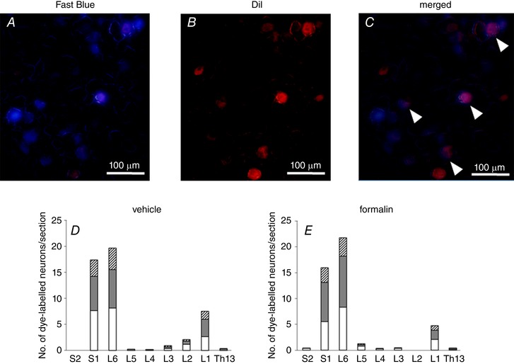
Representative fluorescent microscopic images of DRG neuron labelled with Fast Blue (A) and DiI (B) injected into the bladder wall and the ventral lobes of prostate, respectively. C, a merged image of A and B showed that some neurons were labelled with both dyes, as indicated by arrowheads. Scale bars; 100 μm. The number of neurons dye‐labelled with Fast Blue (white column), DiI (grey column) and both (hatched column) in sections of Th13 to S2 DRG from vehicle‐injected (D) and formalin‐injected rats (E) was quantified (6 DRG from 3 rats per groups). Columns represent the mean number of dye‐labelled neurons per section.