
Figure 5. Recordings of responses to electrical stimulation.
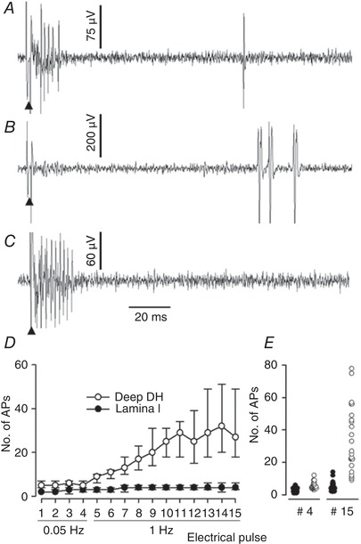
A–C, recordings of different units illustrating the various pattern of innervation of lamina I SPB neurons observed in the present study. Electrical stimulations were delivered with a pair of needles inserted in the receptive field (10 mA, 0.2 ms). A, predominant myelinated fibre innervation and reduced non‐myelinated fibre innervation (typical of nociceptive‐specific neurons in the present study). B, heat‐specific unit receiving input from non‐myelinated fibre only. C, nociceptive‐specific unit receiving input from myelinated fibre only. Arrowhead: electrical stimulation artefact. E, value of the median number of C‐fibre related APs (+75th percentile, −25th percentile) obtained during peripheral electrical stimulations at 0.05 Hz (4 pulses) and then 1 Hz (11 pulses) of lamina I SPB neurons and deep dorsal horn (DH) neuron (n = 23 in each group). Wind‐up of lamina I SPB neuron was noticeably reduced compared to that of neurons located deeper in the dorsal horn. F, dot plot individual values of the number of C‐fibre‐related APs obtained for the 4th pulse (0.05 Hz) and 15th pulse (1 Hz) during the wind‐up test for the lamina I SPB neurons (filled circles) and deep dorsal horn neurons (open circles).