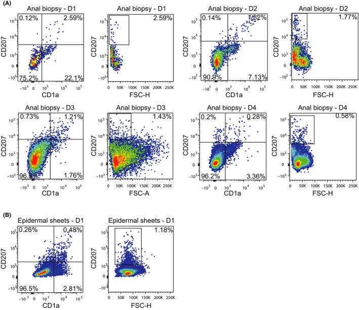
Figure 1. Langerhans cells are present in anal mucosal biopsies from HIV‐1 infected MSM .

(A) Single cell suspensions of anal biopsies from HIV‐1 infected individuals were stained with antibodies against CD45, CD19, CD20, CD56, CD3, CD1a and CD207 and analysed by flow cytometry. The percentage of cells present is depicted in the upper‐right corner of the dot plots. Four representative donors out of six donors is depicted. (B) Single cell suspensions of epidermal sheets were stained with antibodies against CD1a and CD207 and analysed by flow cytometry. One representative donor is depicted. D1, Donor 1; D2, Donor 2; D3, Donor 3; D4, Donor 4.
