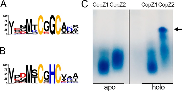
Figure 1.
Structural differences of CopZ1 and CopZ2. Conserved Cu+ binding motifs of (A) CopZ1-like and (B) CopZ2-like proteins. C, native PAGE gel of purified CopZ1 and CopZ2 in the absence (left) and presence of equimolar amounts of Cu+ (right). The vertical dividing line in panel C indicates where the image has been spliced; all signals were from an identical original image and have not been altered. Arrow indicates multimeric structures of CopZ2.

