Figure 7.
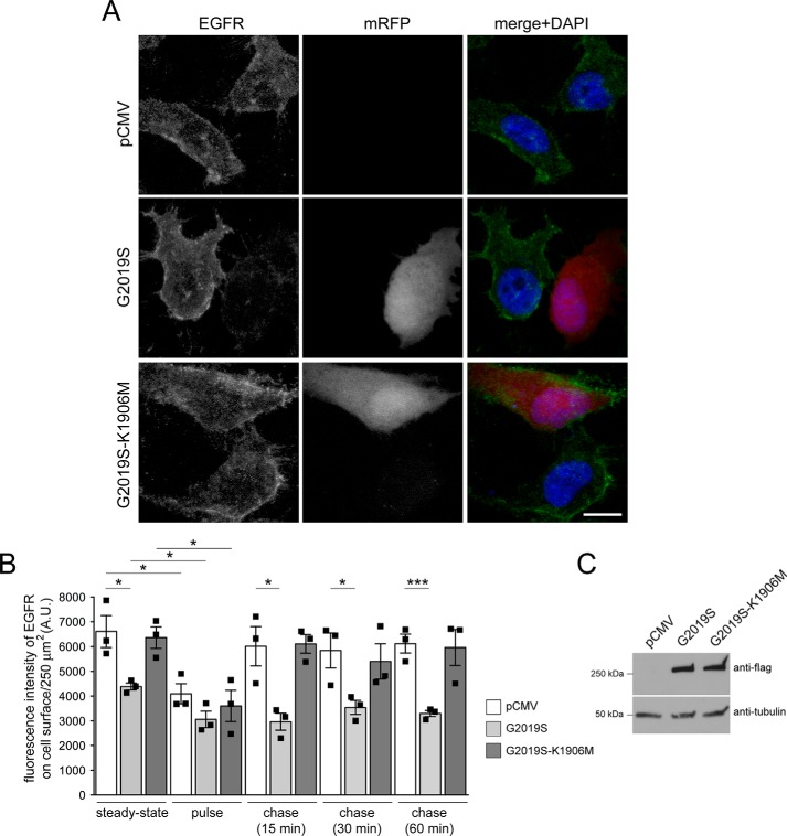
Pathogenic G2019S LRRK2 causes a deficit in EGFR recycling. A, example of HeLa cells transfected with pCMV or cotransfected with mRFP and either G2019S or kinase-inactive G2019S-K1906M LRRK2 and stained with an antibody against the extracellular domain of the EGFR in the absence of permeabilization to visualize only surface EGFR. Scale bar, 10 μm. B, quantification of fluorescence intensity of surface levels of EGFR at t = 0 min (steady-state), upon triggering internalization of the EGFR (pulse), or upon chase for various time points to assess recycling rates (chase) as described under “Materials and methods.” n = 3 independent experiments. *, p < 0.05; ***, p < 0.005. C, HeLa cells were transfected as indicated, and cell extracts (30 μg) were analyzed by Western blotting for FLAG-tagged LRRK2 levels and tubulin as a loading control. A.U., arbitrary units. All error bars represent S.E.M.

