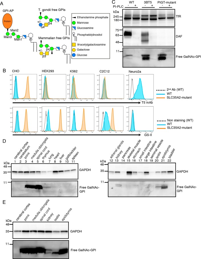
Figure 1.
Detection of free GPIs in some cultured cell lines and mouse tissues using T5 mAb. A, common structure of GPI-AP and structures of free GPIs from T. gondii and mammals. Monosaccharide symbols are according to the Symbol Nomenclature for Glycans (40). B, flow cytometric analysis of wildtype (WT) and SLC35A2 (UDP-Gal transporter)-deficient cell lines. WT (blue) or SLC35A2-mutant (orange) CHO, HEK293, K562, C2C12, and Neuro2a cells were stained with T5 mAb plus secondary antibody for free GPI (top) or with lectin GS-II for nonreducing terminal GlcNAc (bottom). Dotted lines, WT cells stained by secondary antibody only. C, Western blotting of free GPIs of CHO cells. Lysates of WT (3B2A), SLC35A2-mutant (3BT5), and PIGT-mutant cells treated with or without PI-PLC were analyzed by Western blotting. DAF, a GPI-AP; TfR, a loading control. D and E, Western blot analysis of various mouse tissue lysates using T5 mAb. GAPDH was used as a loading control.

