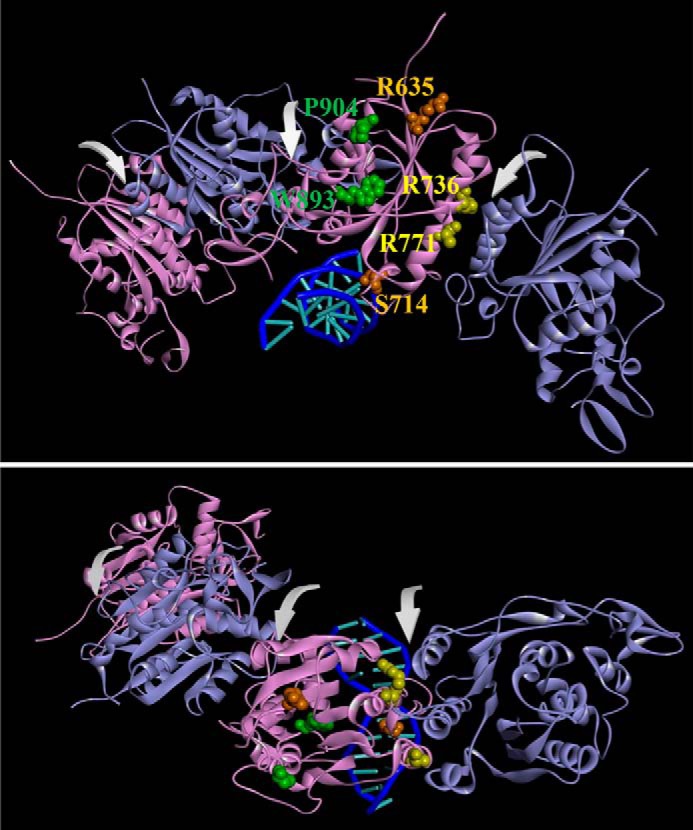
Figure 1.

Mutations from AML patients in a DNMT3A homotetramer model. A model of the DNMT3A homotetramer (alternating purple and cyan monomers) bound to DNA was generated by aligning DNMT3A monomers to DNMT3L in a DNMT3A–DNMT3L heterotetramer crystal structure (PDB ID code 2QRV) followed by a subsequent alignment of a DNMT3A monomer to a M.HhaI-dsDNA co-crystal structure (PDB ID code 3EEO). Arrows in front view (A) and top view (B) indicate dimer and tetramer interfaces. Mutated residues are categorized based on location as follows: surface, orange; tetramer interface, yellow; and internal, green.
