Figure 2.
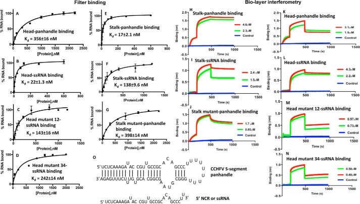
Binding studies performed by filter-binding assay and biolayer interferometry. Binding profiles for the association of stalk domain, stalk mutant domain, head domain, head mutant domain 16, and head mutant domain 34 with CCHFV S-segment vRNA panhandle and ssRNA sequence, generated by filter-binding assay, are shown in A–G. Interaction of each domain with the type of RNA is mentioned inside each panel. Binding results from A–G were also confirmed using biolayer interferometry (BLI). Representative BLI sensograms showing over time association and dissociation of protein with RNA are shown in H–N. The sensograms were generated at two protein concentrations, shown by red and blue in H–N (see “Experimental procedures” for details). Again each panel is internally labeled to show the name of the protein domain and interacting RNA. The dissociation constants (Kd) were calculated as described under “Experimental procedures.” The CCHFV S-segment panhandle and ssRNA used in A–N are shown in O. The panhandle sequence composed of 30 nucleotides from both 5′ and 3′ termini of CCHFV S-segment vRNA, separated by a six-residue uracil loop, was folded by M-fold. It generated the secondary structure, shown in O.

