Figure 3.
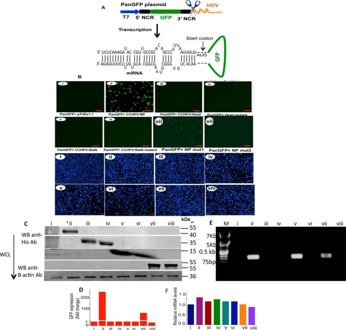
Monitoring the N protein-panhandle binding using an in vivo reporter assay. A, a cartoon showing panGFP plasmid in which GFP (green) flanked by sequences encoding 5′ and 3′ NCR of CCHFV S-segment vRNA (black) was cloned between T7 promoter (blue) and HDV (orange). The mRNA expressed from T7 promoter was folded by M-fold, revealing the formation of panhandle structure by the base pairing of partially complementary nucleotide sequence of the 5′ and 3′ NCR. B, BSRT7.5 cells stably expressing T7 RNA polymerase were seeded in six-well plates and transfected with 2 μg of either empty vector pTriEx1.1 (i), pTriEx-CCHFV-NP plasmid expressing WT CCHFV N protein (ii), pTriExhead plasmid expressing the head domain of the CCHFV N protein (iii), pTriExhead-mutant34 plasmid expressing the head mutant domain 34 (iv), pTriExstalk plasmid expressing the stalk domain of CCHFV N protein (v), pTriExstalk-mutant plasmid expressing the stalk domain mutant (vi), pTriEx CCHFV N protein mut1 plasmid expressing N protein mut1 (vii), or pTriEx CCHFV N protein mut2 plasmid expressing N protein mut2 (viii), as mentioned in the text. Twenty-four hours post-transfection, cells were again transfected with an equal concentration of PanGFP plasmid. Cells were examined under a fluorescence microscope 36 h post second transfection to record the GFP signal (upper eight panels). The experiment was repeated and the cells were stained with DAPI (lower eight panels). The size of the bar in each panel is 100 μm. C, cells from B were lysed and examined by Western blot analysis using anti-His tag antibody to monitor the expression of C-terminally His-tagged fusion proteins from transfected plasmids. D, the cells from B were also examined by FACS. The GFP signal from B (i–viii, upper eight panels) was quantified, normalized related to control (lane 1), and plotted in D. E, the cell lysates from B, containing equal amounts of GFP mRNA, were incubated with Ni-NTA beads. His-tagged proteins bound to washed beads were eluted and total RNA was purified from eluted material. The GFP reporter mRNA in the eluted material was detected using a primer set complementary to the GFP gene. F, total RNA was purified from cell lysates obtained from B. The GFP reporter mRNA was quantified by real time PCR. F shows the equal expression of the reporter GFP mRNA in cells from B.

