Figure 5.
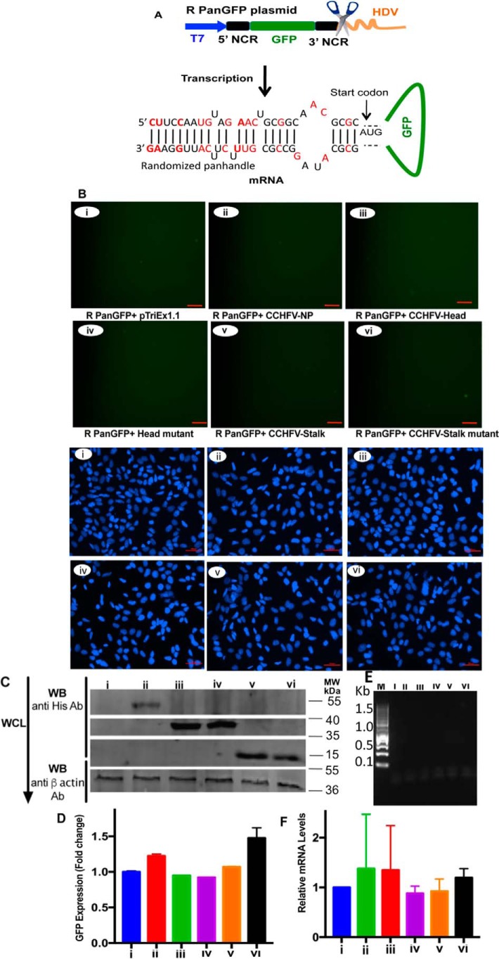
Randomization of 5′ and 3′ NCR sequences inhibited the N protein-panhandle interaction in vivo. A, cartoon showing RpanGFP plasmid expressing a GFP reporter mRNA flanked by randomized sequences of 5′ and 3′ NCR of CCHFV S-segment vRNA. The mutations are shown in red. B, BSRT7.5 cells were transfected with plasmids and examined by fluorescence microscope, except the PanGFP plasmid in Fig. 3B was replaced with RpanGFP plasmid in B. Upper and lower six panels show the GFP expression and DAPI staining, respectively. C, cell lysates from B were examined by Western blot analysis as described in the legend to Fig. 3B. D, cells from B were examined by FACS as described in the legend to Fig. 3D. E, cell lysates from B (upper six panels), containing equal amounts of GFP mRNA, were loaded on Ni-NTA beads and GFP mRNA eluted from washed beads was detected by PCR, as described in the legend to Fig. 3E. F, GFP mRNA in cell lysates from B was quantified by real time PCR exactly as described in the legend to Fig. 3F.

