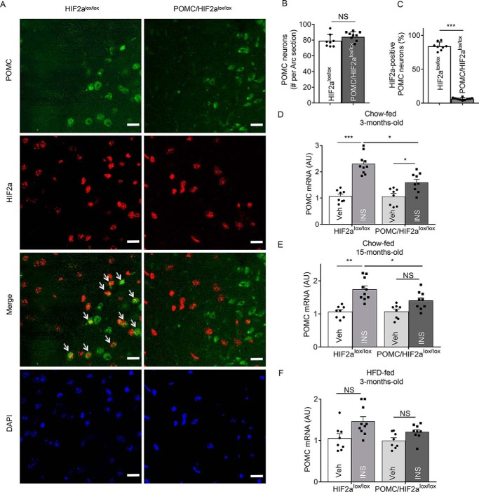
Figure 3.
Role of HIF2α in Pomc neurons in insulin-dependent regulation of Pomc mRNA. A, hypothalamic sections from POMC/HIF2αlox/lox mice and control HIF2αlox/lox mice were stained for HIF2α (red) and POMC (green) in the region containing the arcuate nucleus (Arc). DAPI staining shows the nuclei of cells. Arrows point to POMC neurons that are strongly positive for HIF2α. Scale bar: 25 μm. B and C, POMC neurons (B) and HIF2α-positive population (C) in the Arc sections were counted according to immunostaining. ***, p < 0.001, n = 4 mice per group (biological replicates). Error bars reflect mean ± S.E. D–F, POMC/HIF2αlox/lox mice and HIF2αlox/lox mice at the indicated ages and diet conditions were fasted 24 h and received a third-ventricle injection of insulin (INS) versus vehicle (Veh), and 2 h later the hypothalamus was harvested for the measurement of Pomc mRNA. *, p < 0.05; **, p < 0.01; ***, p < 0.001; n = 8 mice per group (biological replicates). Error bars reflect mean ± S.E. NS, not significant.

