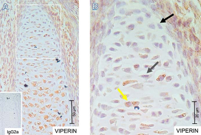
Figure 1.

Viperin expression in the developing embryonal growth plate. 5-μm-thick formalin-fixed paraffin-embedded tissue sections were prepared from embryonic day 15.5 NMRI mouse embryos. Spatiotemporal expression of viperin was detected immunohistochemically. IgG2a was used as an isotype control. A, overview of a representative immunohistochemically stained growth plate with IgG2a negative control in the inset. B, indicated area from A enlarged. The black arrow indicates a representative cell with barely detectable viperin expression; the gray arrow indicates a representative cell with weak viperin expression; and the yellow arrow indicates a representative cell with high viperin expression. The scale bars are indicated for magnification reference.
